By accessing this data, you agree to the terms of the MouseBIRN Data Use Agreement.
Dr. Joanna Jankowsky at Caltech has developed a Tet-off APP model, in which the expression of APP can be controlled with antibiotic treatment. These mice show several significant advantages for use as a model for Alzheimers.
These mice show several significant advantages for use as a model for Alzheimers. Dr. Russ Jacobs at Caltech has collected in vivo and ex vivo high resolution volumes of 16 mice. Recently, higher resolution actively stained specimens were collected at CIVM to examine correlated changes in brain morphology accompanying the production of plaques.
Overview
Alzheimers Disease (AD) is the most common cause of senile dementia in elderly patients. It is characterized by the accumulation of a small peptide known as amyloid beta. While the presence of cognitive decline in 2 or more functional domains suggests the presence of AD, definitive diagnosis can only be made at autopsy. Current treatments for AD based on increasing cholinergic tone provide only temporary slowing of disease progression – staving off cognitive decline by 1 year at most before the disease worsens. Research is underway to enable early diagnosis of AD and to develop new therapies aimed at inhibiting amyloid beta production.
Magnetic resonance (MR) imaging may be especially useful for the diagnosis and treatment of AD. Several studies have been done both in-vivo and ex-vivo using mouse models to characterize the tissue changes in AD progression. Techniques have ranged from direct visualization of plaques to indirect measurement of co-morbid parameters that change with increasing pathology.
Here we aim to develop simple in-vivo/ex-vivo MR protocols that provide high resolution of the neuro-anatomy and amyloid pathology in a mouse model for AD. In-vivo images were analyzed using texture analysis to determine the validity of this technique for use in AD imaging. Ex-vivo images were analyzed by large volume techniques to visualize plaque load across the whole brain. While ex-vivo techniques will benefit the understanding of basic disease mechanisms in animal models of AD, the development of accurate in-vivo image analysis techniques will be critical to the use of MRI in human diagnosis.
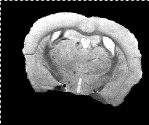
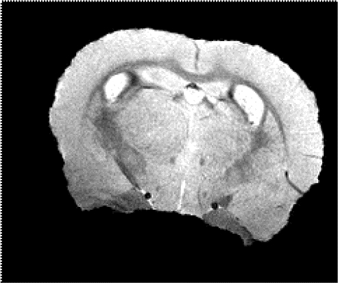
Figure 1. All fifteen mice survived the in-vivo imaging before being euthanized. Representative images of a control mouse and +/+ mouse at 12 months old are shown.
Usage
SHIVA or MBAT are recommended for visualizing the datasets.
Download
The individual data files in analyze format (hdr and img files required for each data set) are contained in the following archives:
exvivo
invivo


